Innovations in Orthopedic Surgery: Breakthroughs and Progress
This article explores key breakthroughs in orthopedic surgery, highlighting minimally invasive techniques like arthroscopy, and advances in imaging technology such as MRI and 3D imaging. It details historical milestones, patient benefits, and the evolution of diagnostic tools that enhance surgical precision. The discussion underscores how technological innovations have improved recovery times and surgical outcomes for various joint and soft tissue conditions, transforming orthopedic care into a more effective and less invasive field.
Innovations in Orthopedic Surgery: Breakthroughs and Progress
Recent advancements in orthopedic medicine reveal that minimally invasive procedures involving fewer tissues lead to lower infection risks and reduced tissue shock, resulting in faster recovery times. These improvements enhance patient outcomes significantly.
Progress in the 20th Century
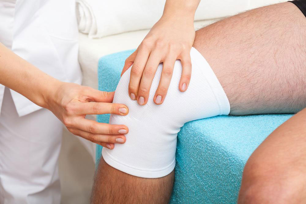
In the early 1900s, limited manufacturing technology hindered the creation of small surgical tools. However, driven by innovation, pioneers like Dr. Masaki Watanabe established arthroscopic surgery by developing the first arthroscope in the 1950s. Dr. Robert W. Jackson later introduced these techniques to the US during the 1960s, initially facing skepticism. Early applications targeted sports injuries like meniscal tears and ligament repairs, with challenges including limited space and instrument maneuverability.
The major patient benefits included less blood loss, decreased pain, quicker recovery, and shorter hospital stays. These advantages sparked widespread acceptance, leading to rapid expansion of arthroscopy into various joints and surgeries. The advent of television monitors further advanced the technique, enabling its use across different joints, including the temporomandibular joint.
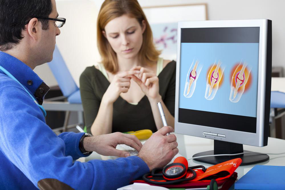
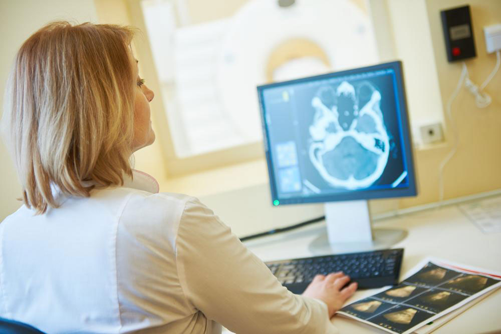
Concurrently, innovations in imaging technology such as MRI and 3D imaging revolutionized orthopedic diagnostics. The improved clarity of ultrasonography enhanced soft tissue visualization, aiding in precise diagnoses of tendons and other structures. These imaging advances allowed detailed surgical planning, resulting in more accurate procedures and better outcomes.
Disclaimer:
The information shared here spans multiple topics, offering valuable insights for readers. The editorial team’s research aims to inform, but articles are not definitive. The website is not responsible for discrepancies or errors across external sources. Additionally, some schemes or offers may not be covered here but could benefit readers more.