Understanding Bone Density Testing: Procedures and Types
Learn about the procedures and types of bone density tests used to diagnose osteoporosis. Discover how DXA and QCT scans work, what results mean, and their importance in early diagnosis. This guide provides essential insights to help you understand bone health assessment methods.

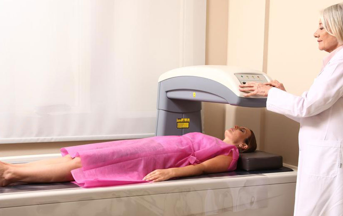
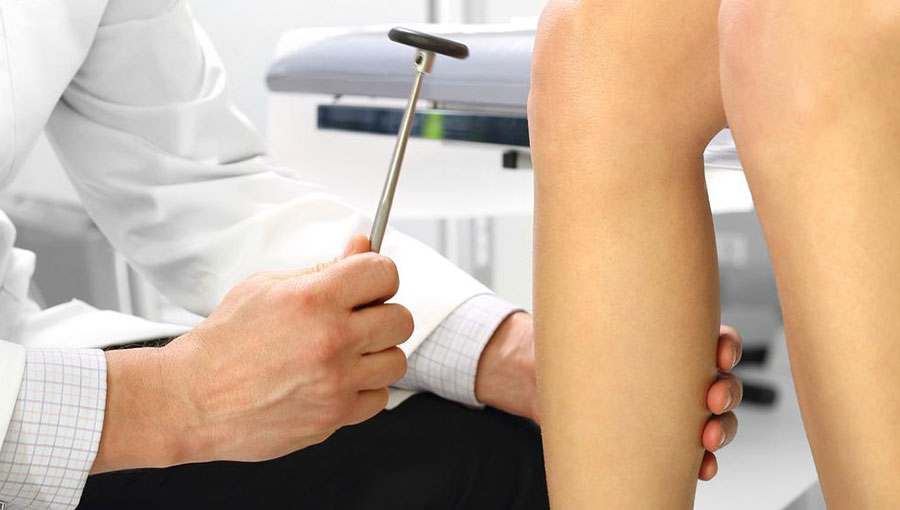
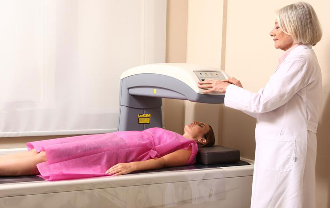
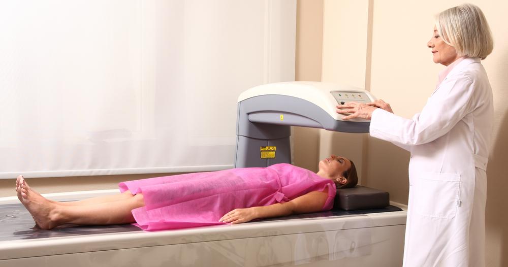
Bone density assessments are essential diagnostic tools that measure the mineral content and strength of bones. These tests are crucial for diagnosing osteoporosis early, often before fractures occur. Typically, scans of the spine or hip are performed, as these are common sites of fractures. Prior to the availability of these tests, osteoporosis was often identified only after a fracture. The procedure involves lying on a padded platform while a small device scans the bones. The radiation exposure is minimal, comparable to a chest X-ray, and the process takes about 10 to 30 minutes.
There are mainly two types of bone density tests. The first, DXA (Dual-energy X-ray Absorptiometry), uses a small dose of radiation to generate images that reveal bone loss. It requires no special preparation, just avoid jewelry and calcium supplements before the scan. The second, QCT (Quantitative Computed Tomography), utilizes a CT scanner to measure bone density, especially useful for individuals with scoliosis or soft bone assessments near the spine. Results are reported as T-scores and Z-scores, which help determine bone health status.
Results interpretation includes:
T-score above -1 indicates normal bone density.
T-score between -1 and -2.5 suggests low bone density, at risk of osteoporosis.
T-score -2.5 or lower confirms osteoporosis.
Z-score indicates how your bone density compares to a healthy peer of the same age and sex; a Z-score below -2 may point to secondary causes of bone loss.
Note: The information provided is for educational purposes and should not replace professional medical advice. Consult a healthcare provider for diagnosis and treatment options.