Understanding Bone Density Testing and Its Importance
This article explains the importance of bone density tests in detecting osteoporosis, highlighting risk factors and the testing process. It underscores the role of scans in monitoring bone health, emphasizing non-invasive methods and the significance of early detection. Suitable for individuals at risk, the article provides essential insights into bone health management and disease prevention, suitable for both patients and healthcare providers. Understanding these tests can lead to timely diagnosis and treatment of osteoporosis, reducing fracture risk and improving quality of life.
Understanding Bone Density Testing and Its Importance
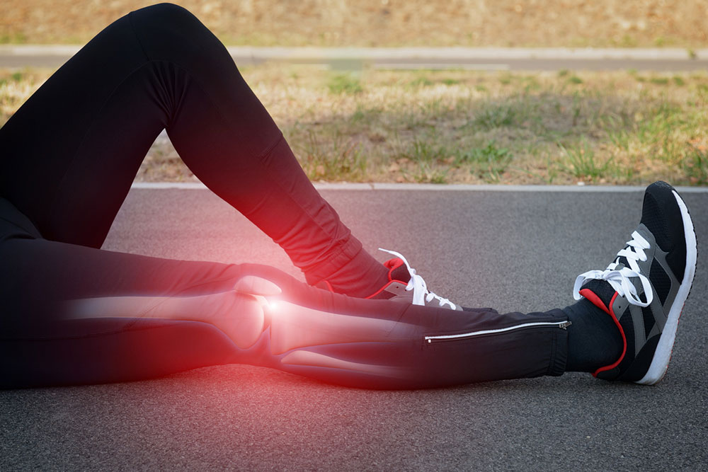
Osteoporosis is a condition characterized by weakened bones, which increases the risk of fractures, especially in the spine, wrist, and hips. Although more common in women, men are also susceptible. In the U.S., approximately 10 million individuals (8 million women and 2 million men) are affected, with around 34 million at high risk due to decreased bone density (osteopenia).
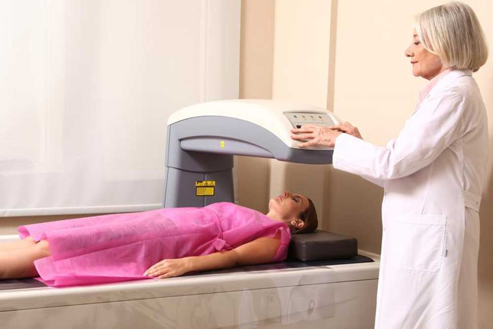
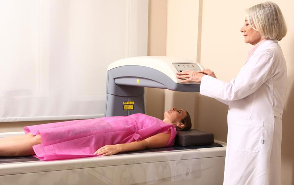
A bone density test is a quick, non-invasive procedure used to detect osteoporosis risk and monitor treatment effectiveness.
Doctors may recommend a bone density scan if:
Height reduction: Losing 1.6 inches or more may indicate spinal fractures linked to osteoporosis.
Broken bones: Fragile bones tend to fracture more easily.
Medication use: Long-term steroid use, such as prednisone, can hinder bone regeneration, raising osteoporosis risk.
Organ transplants: Transplant recipients have higher susceptibility due to immunosuppressants affecting bone health.
Hormonal changes: Postmenopausal women experience lowered estrogen levels, and certain cancer treatments can reduce hormones like testosterone in men, both increasing osteoporosis risk.
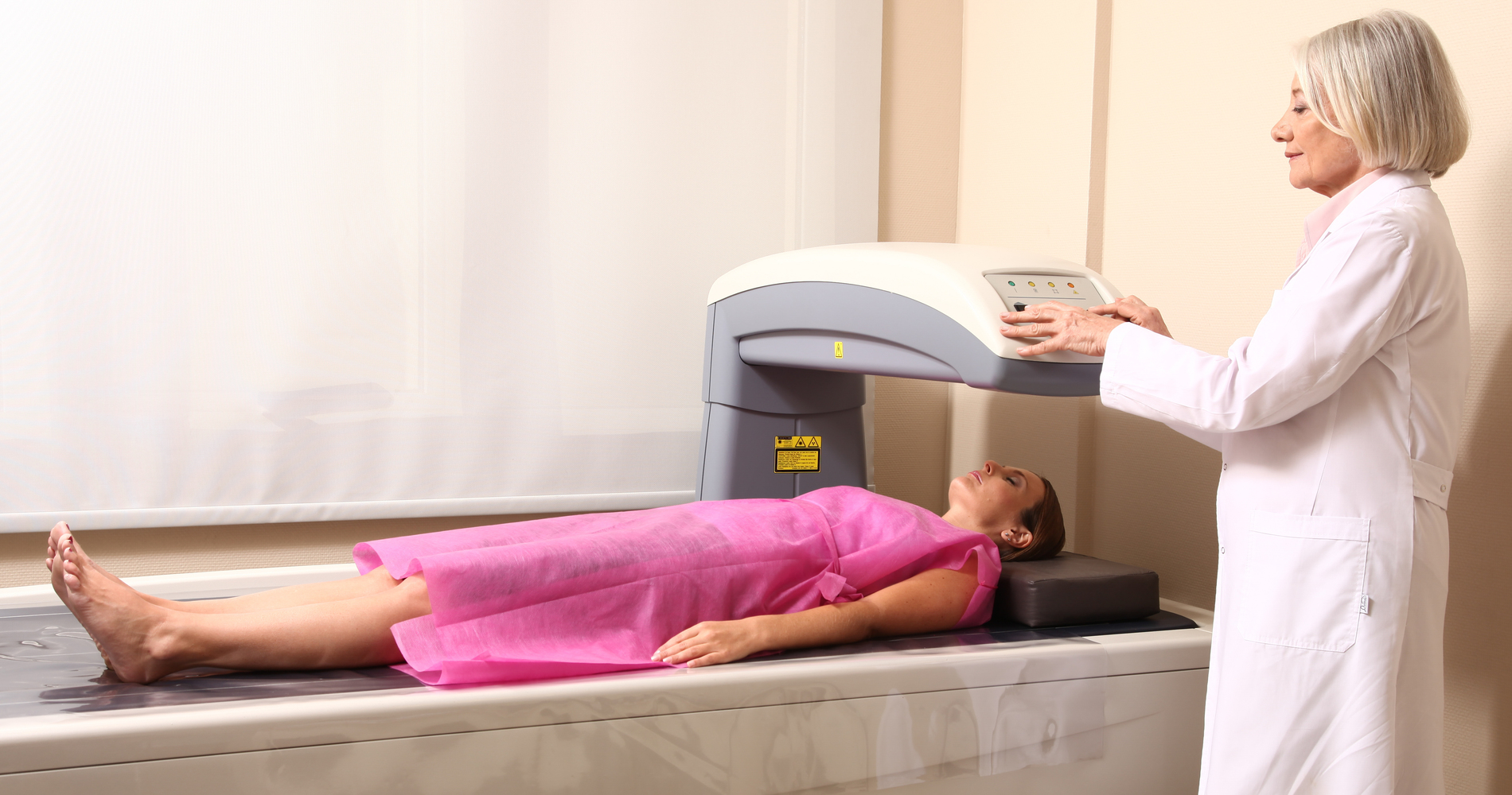
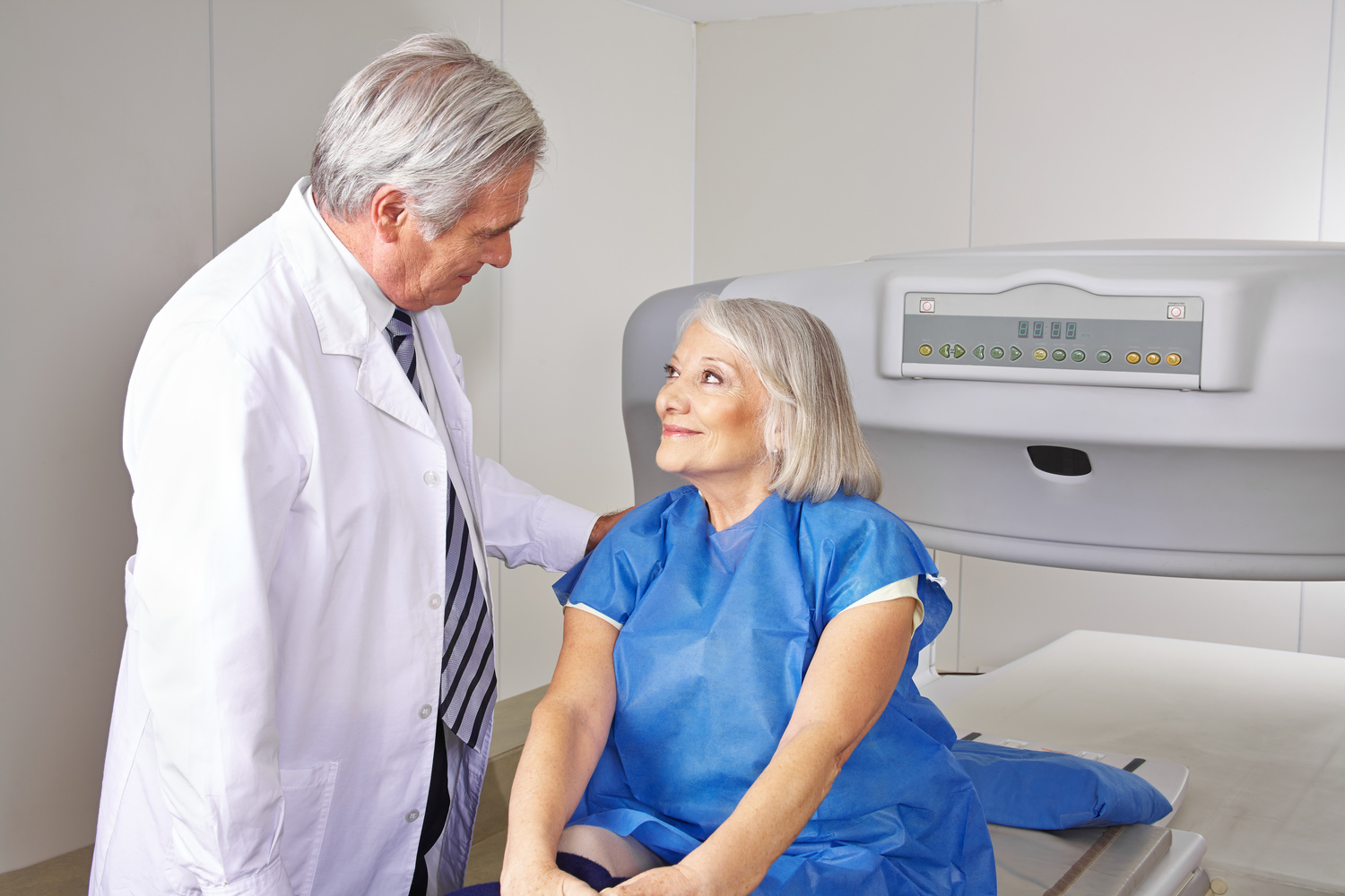
The scan employs a low-dose X-ray to detect signs of decreased bone density and mineral loss. It focuses on the hips and spine, with forearm scans used when hips cannot be evaluated or in hyperthyroidism cases. For individuals under 60, hip scans are usually recommended.
Central machines assess density in the whole body, spine, and hips, while peripheral machines evaluate heel, shinbone, wrist, knee, and finger bones. The scan lasts approximately 15 minutes, with the individual lying on a table for 5-8 minutes. 3D images generated help assess age-related and disease-related bone changes beyond osteoporosis.