Understanding How MRI Scanners Work for Knee Imaging
Discover how MRI scanners operate to produce detailed images of the knee. The article explains preparation, the working mechanism, and the procedure involved in knee MRI scans, highlighting their safety and diagnostic benefits. Ideal for patients seeking insight into MRI imaging processes.

Understanding How MRI Scanners Work for Knee Imaging
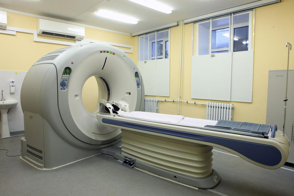
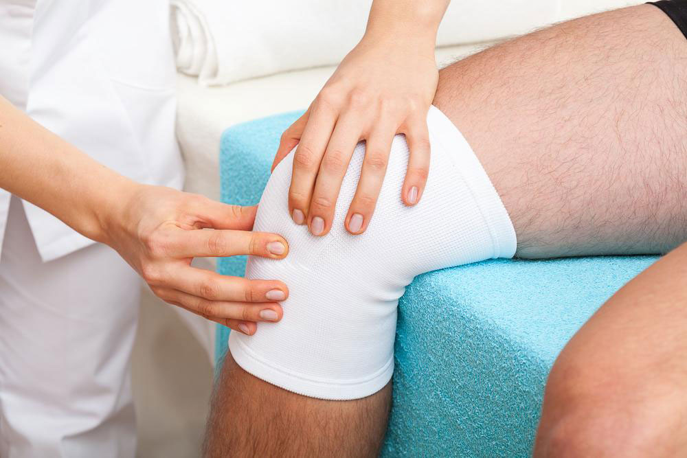
An MRI (Magnetic Resonance Imaging) machine is a donut-shaped device that uses strong magnetic fields and radio waves to generate detailed images of body structures, such as the knee. These scans are valuable for identifying pain, swelling, or inflammation in the knee joint. Unlike X-rays, MRIs do not utilize ionizing radiation, making them a safer choice for detailed internal imaging and helping determine if surgical intervention is needed.
Preparing for a Knee MRI
Patients may be asked to wear a loose gown or clothing without metal fasteners during the scan.
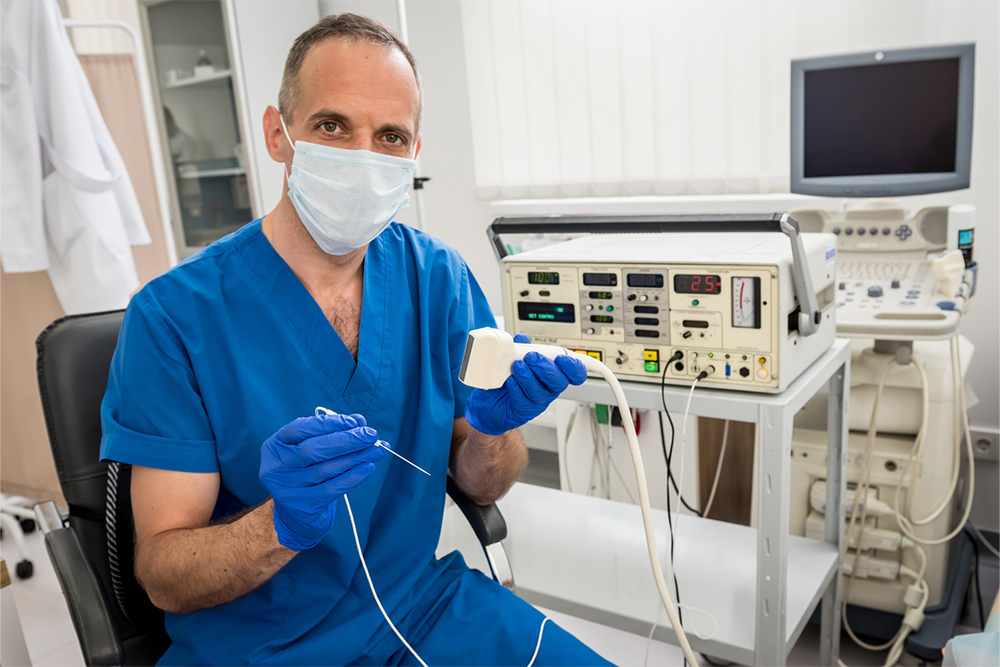
In some cases, contrast agents administered intravenously enhance image clarity, especially for detailed examination of knee tissues.
The radiologist may inquire about allergies before administering contrast substances. For those allergic to iodine, gadolinium might be used as an alternative contrast agent, requiring patient consent.
How MRI Works
The MRI scanner sends radiofrequency pulses that temporarily realign hydrogen atoms within the body. These atoms emit signals based on their tissue environment, which the machine detects to create detailed images. These images can be viewed from various angles, helping identify abnormalities with higher contrast compared to other imaging techniques.
The entire knee MRI procedure can take between 15 to 45 minutes, depending on the complexity. Patients are positioned on a movable table, with straps used to keep the knee steady. Small radio wave emitters are placed near the knee to improve image quality. Most scans are non-invasive and painless, though some patients may feel discomfort or claustrophobia, for which sedation can be arranged.
Note: Our website provides general information on various topics. While we strive for accuracy, readers should consult healthcare professionals for personalized advice. Content is for informational purposes and should not replace expert medical recommendations. The site may not include all available schemes or offers.