Understanding Bone Density Assessments and Their Importance
This article explores the importance of bone density testing in diagnosing osteoporosis. It explains how the procedure works, who should get tested, and what risk factors contribute to bone thinning. Bone health is crucial for preventing fractures, and early assessment can guide treatment plans. The article emphasizes the significance of scans in detecting osteoporosis and monitoring progression or response to therapy, highlighting symptoms, risk factors, and the scanning process for effective bone health management.
Understanding Bone Density Assessments and Their Importance
Osteoporosis is a condition characterized by weakened bones, increasing the risk of fractures, especially in the spine, wrist, and hip. While it predominantly affects women, men are also susceptible. In the U.S., approximately 10 million individuals (8 million women and 2 million men) live with osteoporosis, and an additional 34 million are at high risk due to decreased bone mass, known as osteopenia.
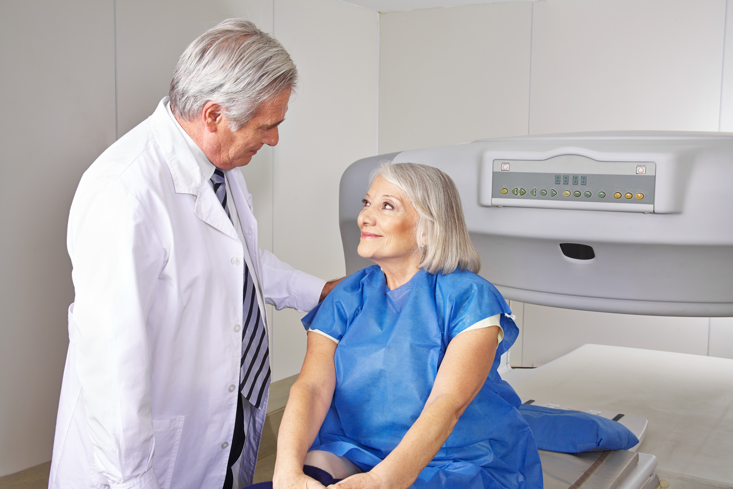
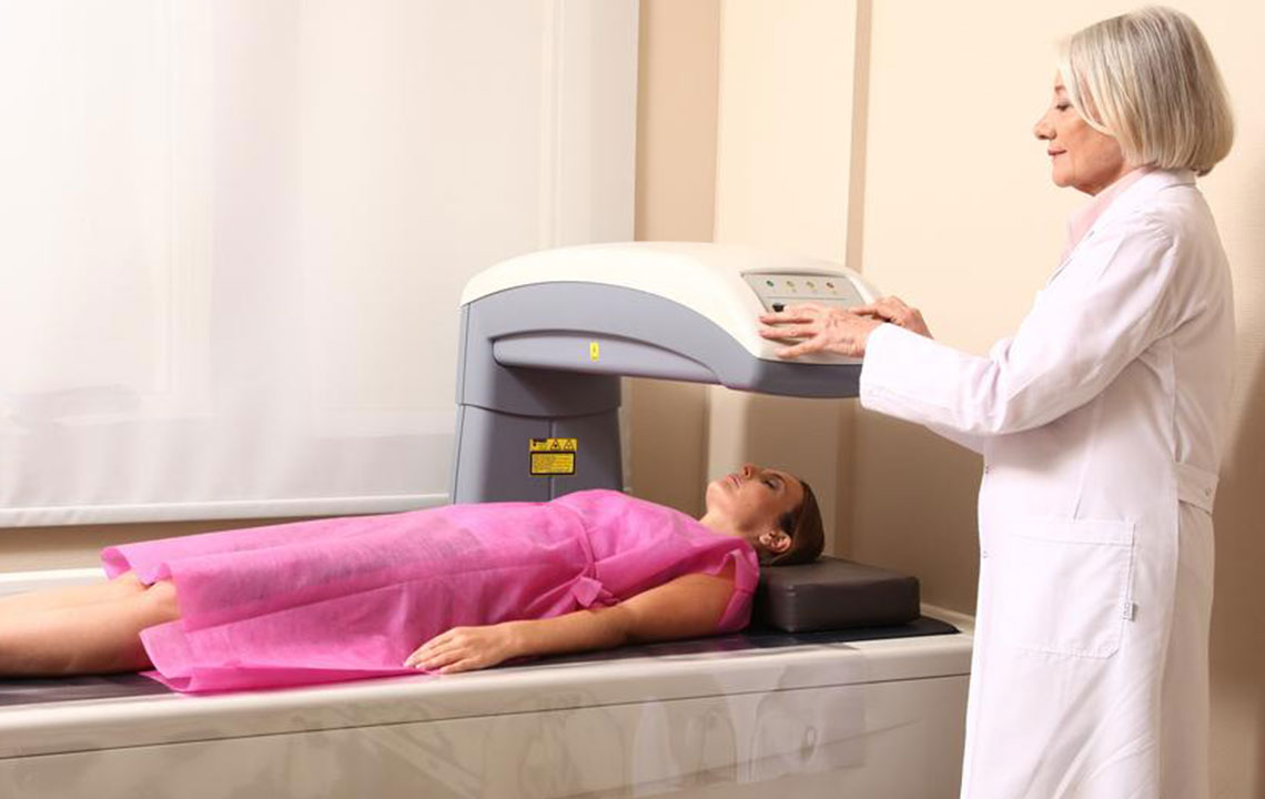
A bone density test is a quick, non-invasive procedure used to diagnose osteoporosis and assess fracture risk. It also monitors treatment effectiveness. Healthcare providers may recommend this test in specific situations.
Height reduction: Loss of over 1.6 inches can indicate spinal fractures due to osteoporosis.
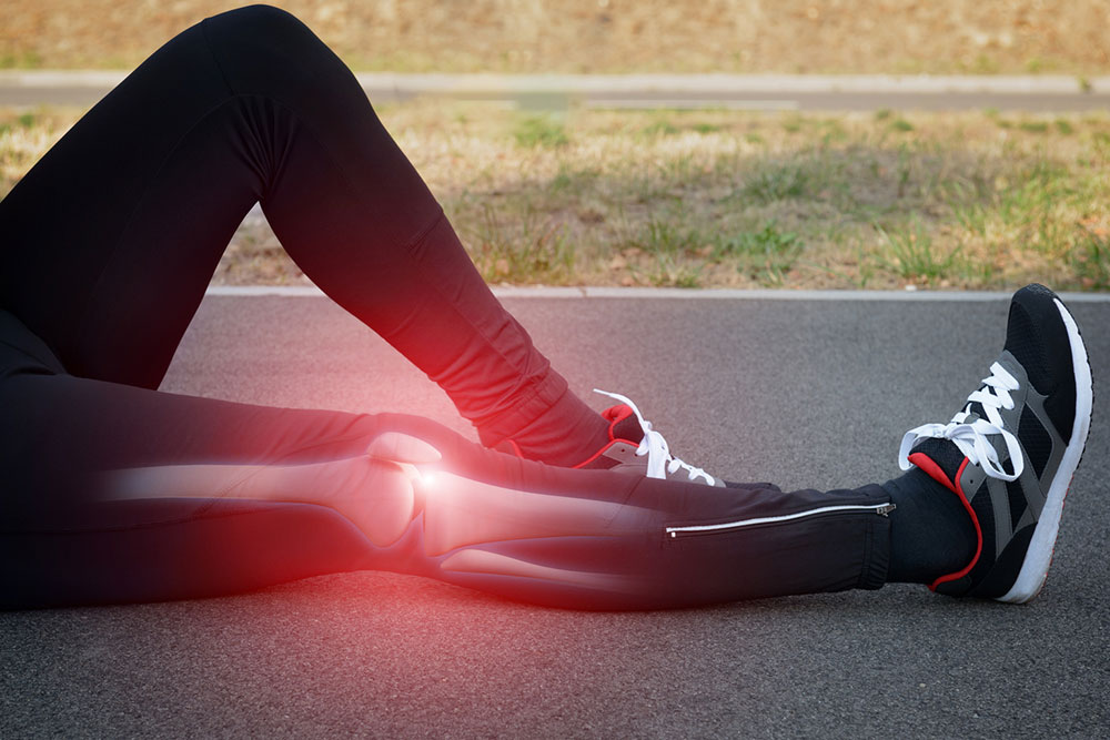
Bone fractures: Fragile bones are more prone to breaking.
Specific treatments: Long-term use of certain medications can hinder bone regeneration, leading to osteoporosis.
Organ transplants: Transplant procedures may increase osteoporosis risk by affecting bone health.
Hormonal changes: Postmenopausal women experience a drop in estrogen levels, while cancer treatments and prostate cancer therapies can lower testosterone, raising osteoporosis risk in men.
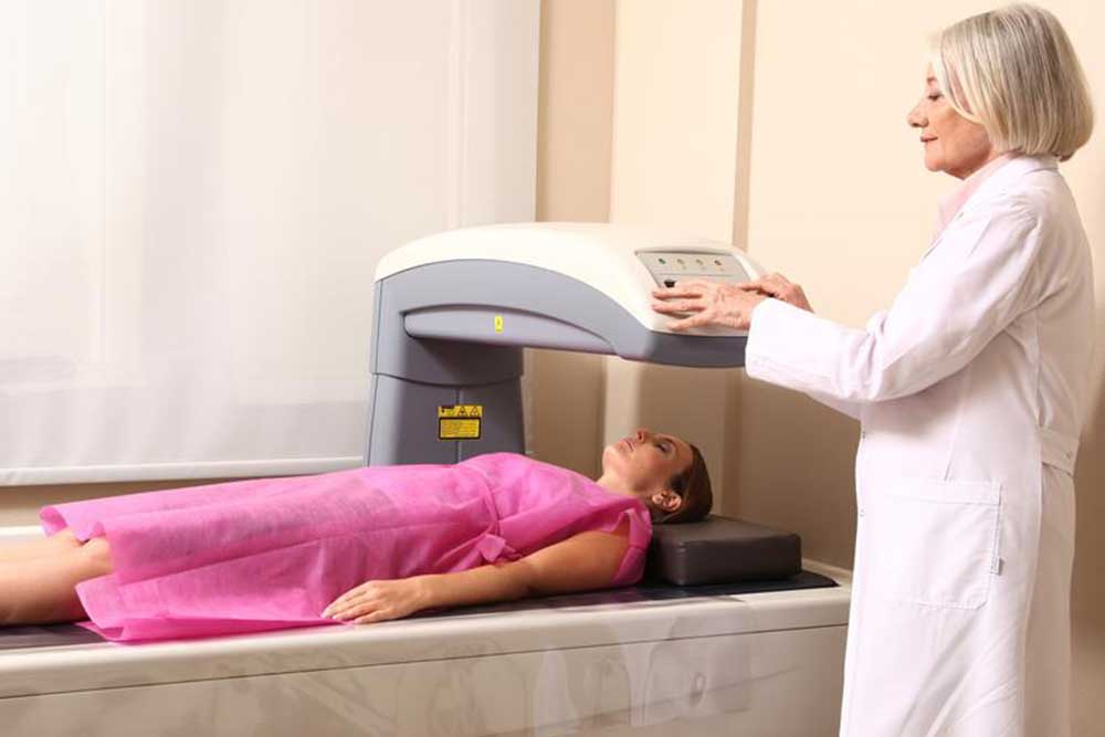
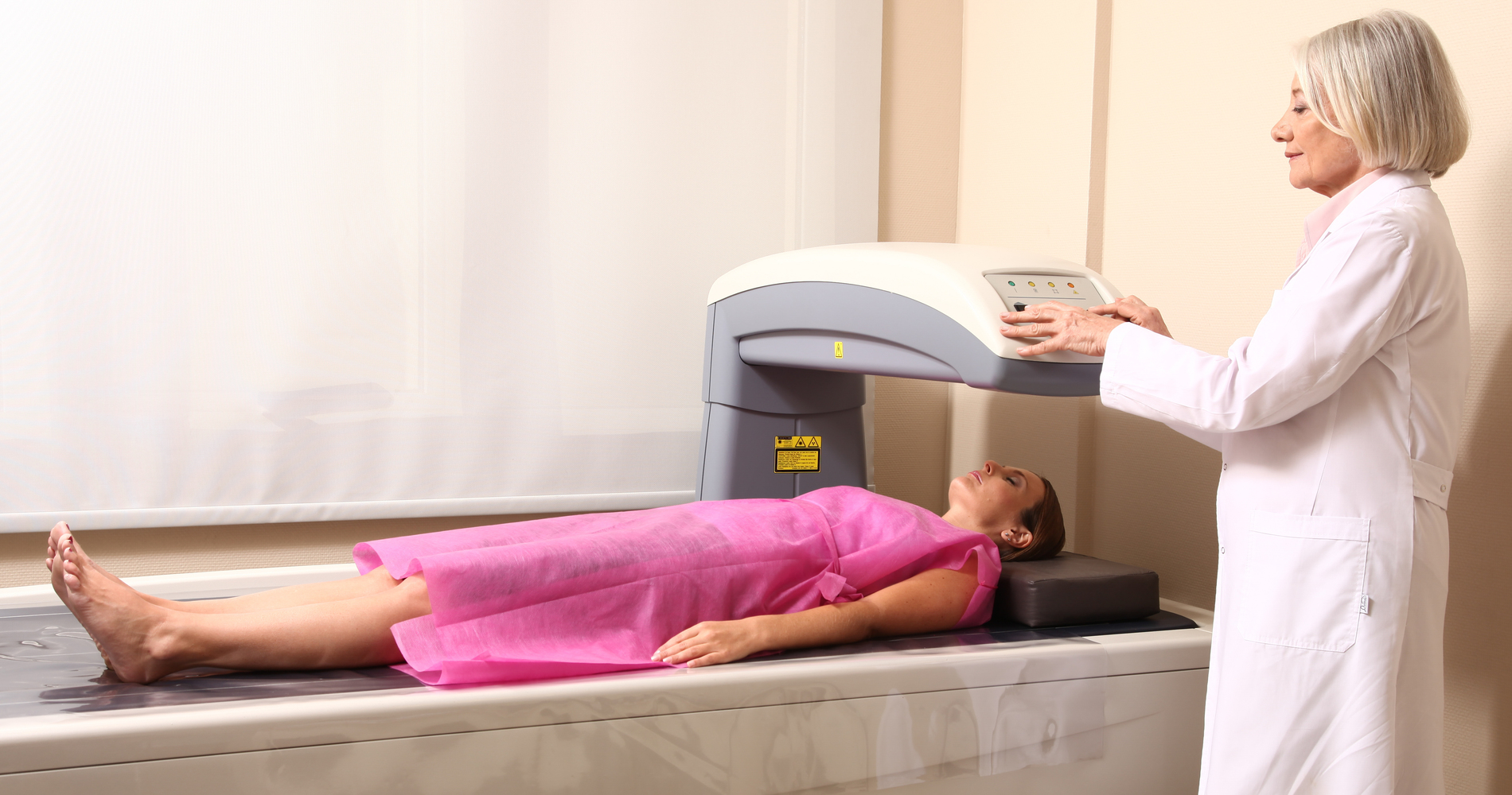
Low-dose X-ray scans detect decreased bone density and mineral loss, focusing on the hips and spine. If the hip cannot be scanned, forearm scans are used, especially for hyperthyroidism patients. For those under 60, a hip scan is typically recommended.
These scans evaluate bone density in the total body, spine, and hips, while peripheral devices measure density in the heel, shin, kneecap, wrist, and fingers. The procedure lasts about 15 minutes, with the person lying on a table for 5-8 minutes. Detailed 3D images help assess age-related changes and bone conditions beyond osteoporosis.