Comprehensive Guide to Early Cancer Screening Tests
A detailed overview of key early cancer detection methods, including imaging, biopsy, ultrasound, and endoscopy. This guide helps patients understand screening options for timely diagnosis and better outcomes, emphasizing the importance of adhering to recommended guidelines and consulting healthcare professionals.
Different Methods for Detecting Cancer Early
Detecting cancer at an early stage is crucial for effective treatment and improved survival rates. Early screening not only aids in timely diagnosis but also minimizes patient discomfort and reduces healthcare costs. The American Cancer Society provides essential guidelines for cancer screening, which healthcare providers and patients should be familiar with. Below are some of the primary tests recommended for early cancer detection.
Imaging Techniques
Imaging examinations allow physicians to visualize internal body structures. Techniques such as ultrasound, X-rays, and advanced imaging help assess the presence of tumors, their size, and spread. These methods are instrumental in determining whether a growth is malignant and in planning appropriate treatments.
Computed Tomography (CT) Scans
CT scans are non-invasive and painless procedures that produce detailed cross-sectional images of bones, organs, and tissues. Using X-ray beams from multiple angles, they help identify tumor size, location, and structure. Additionally, CT scans are useful for guiding treatments like radiofrequency ablation, which destroys tumors with heat.
Magnetic Resonance Imaging (MRI)
MRI employs magnetic fields and radio waves to generate detailed images of soft tissues and internal organs. Sometimes enhanced with contrast dyes like gadolinium, MRI is particularly valuable for detecting cancer spread and pinpointing the origin. Patients lie on a table inside a cylindrical magnet during the scan, which helps in accurate diagnosis.
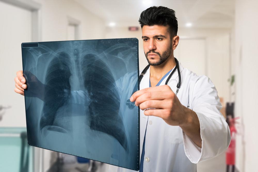
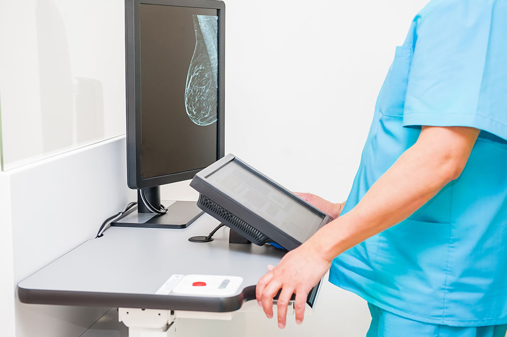
X-ray and Contrast Procedures
X-rays and contrast studies assist in early cancer detection by producing shadow images of bones and internal organs. While less detailed than MRI or CT, they are quick and cost-effective. Mammograms, a specialized breast X-ray, are vital for breast cancer screening. Contrast agents like barium improve visualization of organs such as the gastrointestinal tract, helping identify abnormalities.
Nuclear Medicine Imaging
This technique involves radioactive substances called radionuclides that emit low-dose radiation to create functional images based on tissue activity. Areas affected by cancer absorb differing amounts of these substances, allowing for tumor localization. Although helpful in identifying certain cancers, nuclear scans are often followed by other tests for detailed analysis.
Ultrasound Imaging
Ultrasound uses high-frequency sound waves to produce real-time images of organs and blood flow. It is especially useful for detecting tumors that are not clearly visible on X-rays and differentiating cysts from solid growths. Since it does not involve radiation, ultrasound is safe and rapid, though limited in penetrating air or bones.
Endoscopy Procedures
Endoscopy involves inserting a flexible tube equipped with a camera and light to examine internal organs directly. It is beneficial for diagnosing and treating certain cancers, such as colorectal cancer or esophageal tumors. Therapies including laser ablation and photodynamic treatment can be performed during endoscopy.
Biopsy
A biopsy is performed after initial examinations to confirm cancer diagnosis. It involves removing tissue samples through various techniques like fine-needle aspiration, guided by imaging. The tissue is then analyzed microscopically to determine whether it is cancerous and to assess tumor characteristics.
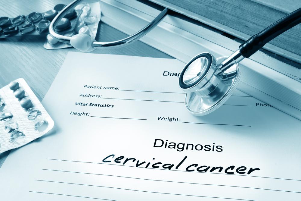
Cytology Tests
Cytology involves examining individual cells collected from tissue scrapes, brushings, or body fluids such as urine and sputum. These tests are useful in diagnosing cancers and other conditions like infections, fetal abnormalities, or Pap smears for cervical cancer screening.