Understanding Catheter-Enhanced Thrombolysis for Deep Vein Thrombosis
Explore the essentials of catheter-assisted thrombolytic therapy for deep vein thrombosis. This minimally invasive procedure utilizes guided catheters to dissolve blood clots, restore circulation, and reduce recovery time. Learn about preparation, procedure steps, equipment used, interpreting results, and potential risks. Suitable for patients seeking effective, less invasive DVT treatment options with fewer hospital stays and quick recovery.

Understanding Catheter-Enhanced Thrombolysis for Deep Vein Thrombosis
Overview of catheter-assisted thrombolytic therapy
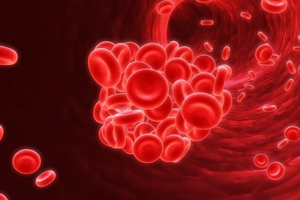
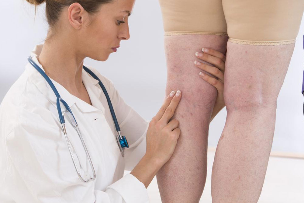
Deep vein thrombosis (DVT) occurs when a blood clot forms in a deep vein, typically in the legs or thighs. Signs include skin redness, swelling, and tenderness along the affected vein. It is more common in individuals over 50 or those with impaired blood flow due to health conditions.
What is catheter-assisted thrombolytic therapy?
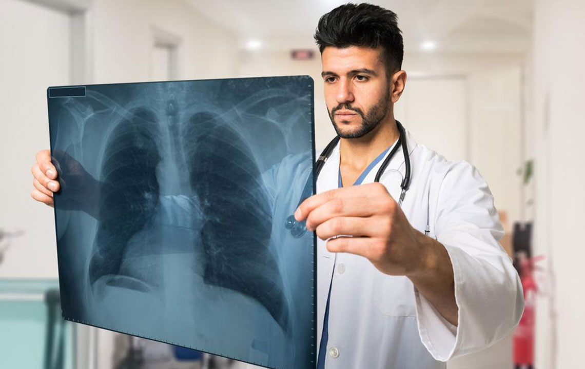
This treatment uses a catheter guided by X-ray imaging to dissolve blood clots within veins.
It restores normal blood flow, protecting tissues and organs from damage caused by blockages.
Advantages of this procedure
It is generally a safe and minimally invasive method for DVT management.
It effectively improves circulation without the need for open surgery.
No surgical incision is necessary, reducing recovery time.
Patients can often avoid extended hospital stays associated with traditional surgeries.
Preparation tips before treatment
Share your current and past medication details with your doctor.
Inform your healthcare provider of pre-existing health conditions.
If pregnant or planning pregnancy, notify your doctor.
Blood tests may be scheduled to assess kidney function and clot status.
Your physician may suggest medication adjustments or additional preparatory steps.
Steps involved in the procedure
Using contrast dye and X-ray guidance, the targeted vein is located.
A small skin puncture is made to insert a catheter into the vessel.
The catheter is carefully advanced to the blood clot site.
Clot removal involves administering medication directly or using devices to break up the clot.
Necessary equipment
A slender plastic catheter, comparable to a spaghetti strand, is used.
X-ray equipment, medication delivery systems, monitors, ultrasound devices, and IV lines are integral to the process.
The specific tools may vary based on the treatment approach prescribed by your doctor.
Interpreting treatment results
An interventional radiologist will evaluate the success of the procedure.
Follow-up treatments may be needed if tissue damage occurred during therapy.
Additional tests, including blood work and imaging, may be conducted during follow-up visits.
Potential risks and considerations
Possible infection risk, approximately 1 in 1,000 cases.
Rare allergic reactions or vessel injury.
Bleeding complications at various sites.
Individuals with kidney issues may experience rare kidney damage.
Clot fragments might migrate, necessitating further intervention.

